Veterinary Diagnostic Lab
At Sun Valley Animal Center, we have advanced tools to help diagnose your pet's medical issues. We offer a variety of services, from digital radiology to ultrasound and cold lasers.
We use radiographic imaging that allows us to produce highly detailed images of your pet's internal structures.
With our diagnostic imaging capabilities, we can efficiently produce diagnostic information about your pet's condition and provide immediate treatment options.

In-House Lab & Veterinary Pharmacy in Ketchum
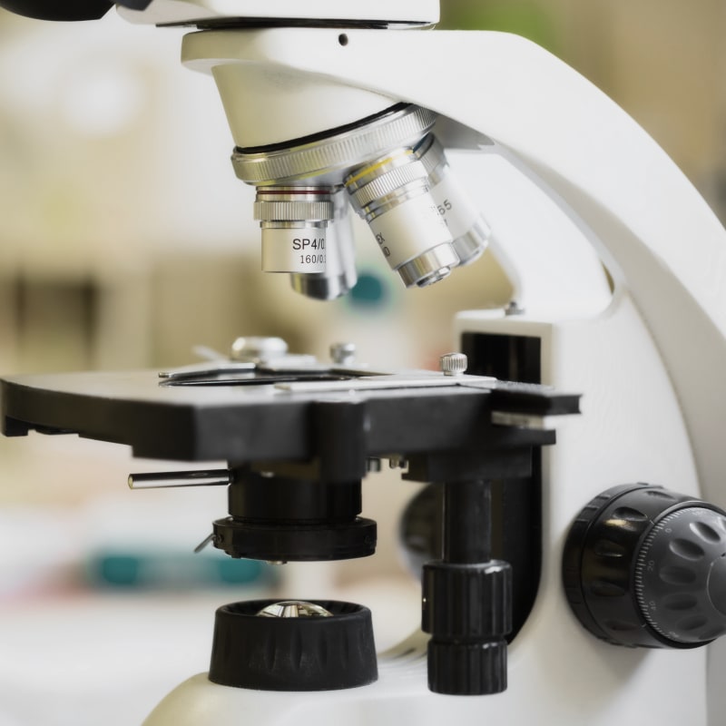
We perform tests and get results quickly in our in-house laboratory so that we can diagnose your pet's symptoms and begin treatment as soon as possible.
Our pet pharmacy in Ketchum is stocked with a range of prescription diets and medications, providing us with quick access to any medications your pet may need while in our care.

Our Diagnostic Services
With our in-house veterinary diagnostics lab we are pleased to offer advanced diagnostic testing to allow our vets to provide rapid diagnosis of your pet's medical issues.
-
Radiography (Digital X-rays)
Using a radiograph (digital x-ray), we can examine your pet's internal systems to reveal information that may be invisible from the outside.
Radiography is safe, painless and non-invasive. It uses only very low doses of radiation. Because the level of radiation exposure required to perform radiography is very low, even pregnant females and very young pets can undergo this procedure.
Radiographs can be used to evaluate bones and organs, and diagnose conditions including broken bones, chronic arthritis, bladder stones, spinal cord diseases and some tumors. -
Myelography
A myelogram is an animal x-ray test in which dye is injected directly into the spinal canal to help show places where disc material in the back may be pinching the spinal cord.
Our veterinary hospital also uses this procedure to help diagnose back and leg pain problems, especially if we anticipate the need for surgery. -
Penn-HIP
Penn-HIP, owned and operated by the University of Pennsylvania, incorporates a method of evaluating the integrity of the canine hip, using multiple disciplines, including biomechanics, orthopedics, clinical medicine, radiology, epidemiology, and population genetics.
The purpose of the program is to direct appropriate breeding strategies aimed at reducing the frequency and severity of osteoarthritis and hip dysplasia in canines.
Dr. Randy Acker has found PennHIP to be extremely informative in his orthopedic cases and considers it to be a more thorough method for assessing breeding soundness. -
Ultrasound
The use of diagnostic imaging allows our team of veterinary professionals to create extremely detailed images of your pet's internal structures.
With ultrasound imaging, we expose part of the body to high-frequency sound waves to produce images of the inside of the body.
Because we capture ultrasound images in real-time, we can see the structure and movement of your pet's internal organs, as well as blood flowing through the blood vessels.
Having this valuable technology available to our vets in our in-house lab means that your dog or cat's condition can be diagnosed quickly and treatment can start sooner. -
CT Scan
CT (computed tomography) Scans combine X-Ray images and computer technology to help us identify and diagnose numerous diseases and disorders in your pet's body. They have become an essential imaging tool used in veterinary medicine.
-
OFA Hips & Elbows
OFA (Orthopedic Foundation for Animals), formed by John M. Olin in 1966 also assesses Canine Hip Dysplasia (CHD) and Degenerative Joint Disease (DJD).
OFA raises money for research to advise, encourage and establish control programs to lower the incidence of orthopedic and genetic diseases in animals. OFA’s traditional method for determining CHD and DJD relies on the standard technique of radiographic positioning of the pelvis or hips only in the extended position, therefore the passive hip laxity is not included in their assessment.
-
Arthrogram
An Arthrogram is a series of images, often x-rays, of a joint following injection of a contrast medium.
-
Gastrointestinal Diagnostics
Gastrointestinal problems are some of the more common complaints in domestic animals, encompassing a large range of problems, from intestinal parasites to cancer.
Diagnostics always start with a good history and physical exam. Further diagnostics can include fecal studies, blood work, radiography, barium studies, and potentially surgery.
-
Laparoscopy
Minimally invasive surgical techniques are seemingly more common in the veterinary world. Just like in people, these procedures are less painful and allow for quicker recovery.
Commonly performed procedures in dogs include ovariohysterectomy, liver and kidney biopsies, as well as gastropexies.
-
Digital Dental X-Rays
If your cat or dog is suffering periodontal disease, much of this damage occurs below the gum line where it can't be easily seen. Digital dental X-Rays help our veterinarians assess your pet's oral health.
Digital X-Rays are safer for your pet. They allow our team of veterinary professionals to examine roots, bones and internal anatomy of your cat or dog's teeth.
With digital X-Rays, the risk of radiation exposure for your pet is significantly lower than with traditional X-Ray technology. We are able to see below the surface of your pet's gum line to fully evaluate each tooth.
This technology allows your Ketchum vet to see results immediately, then project them onto a computer screen to review. -
ECG/EKG
If your veterinarian performs a physical examination and suspects your pet may have a heart disorder, we usually take chest X-Rays and an electrocardiogram (ECG / EKG).
This procedure can be completed easily and quickly. It reveals data that may be integral to your pet's diagnosis. In other cases, a cardiac ultrasound may be required to identify disorders in the chambers of the heart.
